Having bradycardia (say “bray-dee-KAR-dee-uh”) means that your heart beats very slowly. For most people, a heart rate of 60 to 100 beats a minute while at rest is considered normal. If your heart beats less than 60 times a minute, your doctor may diagnose bradycardia.
 A slow heart rate is sometimes normal and can be a sign of being very fit. Healthy young adults and athletes often have heart rates of less than 60 beats a minute.
Bradycardia is caused by something that disrupts the normal electrical impulses controlling the rate of your heart’s pumping action. Many things can cause or contribute to problems with your heart’s electrical system, including:
- Heart tissue damage related to aging
- Damage to heart tissues from heart disease or heart attack
- High blood pressure (hypertension)
- Heart disorder present at birth (congenital heart defect)
- Infection of heart tissue (myocarditis)
- A complication of heart surgery
- Underactive thyroid gland (hypothyroidism)
- Imbalance of electrolytes, mineral-related substances necessary for conducting electrical impulses
- Obstructive sleep apnea, the repeated disruption of breathing during sleep
- Inflammatory disease, such as rheumatic fever or lupus
- Hemochromatosis, the buildup of iron in organs
- Medications, including some drugs for other heart rhythm disorders, high blood pressure and psychosis
These causes are best divided into cardiac and noncardiac causes. Noncardiac causes are usually secondary, and can involve recreational drug use or abuse; metabolic or endocrine issues, especially in the thyroid; an electrolyte imbalance; neurologic factors; autonomic reflexes; situational factors such as prolonged bed rest; and autoimmunity. Cardiac causes include acute or chronic ischemic heart disease, vascular heart disease, valvular heart disease, or degenerative primary electrical disease. Ultimately, the causes act by three mechanisms: depressed automaticity of the heart, conduction block, or escape pacemakers and rhythms.
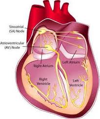 Generally, two types of problems result in bradycardias: disorders of the sinoatrial node (SA node), and disorders of the atrioventricular node (AV node).
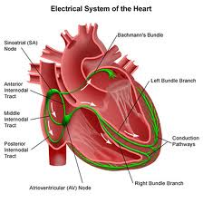
Electrical circuitry of the heart
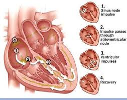
Your heart is made up of four chambers — two upper chambers (atria) and two lower chambers (ventricles). The rhythm of your heart is normally controlled by a natural pacemaker — the sinus node — located in the right atrium. The sinus node produces electrical impulses that initiate each heartbeat.
From the sinus node, electrical impulses travel across the atria, causing the atria to contract and pump blood into the ventricles. The electrical impulses then arrive at a cluster of cells called the atrioventricular node (AV node).
The AV node transmits the signal to a specialized collection of cells called the bundle of His. These cells transmit the signal down a left branch serving the left ventricle and a right branch serving the right ventricle. When the electrical impulse travels down these branches, the ventricles contract and pump blood — the right ventricle sending oxygen-poor blood to the lungs and the left ventricle sending oxygen-rich blood to the body.
Bradycardia occurs when electrical signals slow down or are blocked.
Sinus node problems
Bradycardia often starts in the sinus node. A slow heart rate may occur because the sinus node:
- Discharges electrical impulses at a slower than normal rate
- Pauses, or fails to discharge at a regular rate
- Discharges an electrical impulse that’s blocked before causing the atria to contract
In some people the sinus node problems may result in alternating slow and fast heart rates (bradycardia-tachycardia syndrome).
Heart block (atrioventricular block)
 Bradycardia may also occur because electrical signals transmitted through the atria aren’t transmitted to the ventricles (heart block, or atrioventricular block). The disruption of the electrical signal may occur in the AV node, the bundle of His, or somewhere along the left and right branches that transmit electrical signals to the ventricles. Heart blocks are classified based on the degree to which signals from the atria reach your heart’s main pumping chambers (ventricles).
- First-degree heart block. In the mildest form of heart block, all electrical signals from the atria reach the ventricles, but the signal is slowed down slightly. First-degree heart block rarely causes symptoms and usually needs no treatment if there’s no other abnormality in electrical signal conduction.
- Second-degree heart block. In second-degree heart block, not all electrical signals reach the ventricles. Some beats are “dropped,” resulting in a slower and sometimes irregular rhythm.
- Third-degree (complete) heart block. In third-degree heart block, none of the electrical impulses from the atria reaches the ventricles. When this happens, the bundle of His or other tissues of the ventricles function as a substitute pacemaker for the ventricles. These substitutes result in slow and sometimes unreliable electrical impulses to control the beat of the ventricles.
- Bundle branch block. The interruption of an electrical signal somewhere in the right or left bundle branches — near the end of the pathway for electrical impulses — is called a bundle branch block. The seriousness of bundle branch block depends on whether both branches are affected, the presence of other types of heart block and the degree of damage to heart tissue.