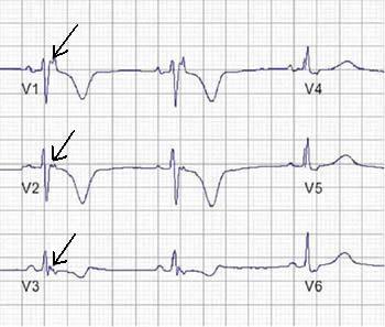
Adenosine thallium scan: A method of examining the heart to obtain information about the blood supply to the heart muscle. Special cameras take a series of pictures of the heart. Radioactive thallium is injected into the bloodstream and serves as a tracer. The tracer attaches to certain cells and makes them visible to the special camera. The tracer attaches to the muscle cells of the heart so the imaging camera can take pictures of the heart muscles. If an area of the heart does not receive an adequate flow of blood, the cells in the underserved area do not receive as much tracer and it appears as a darker area on the picture taken by the camera. Continue reading

Adenosine thallium scan
Leave a reply
