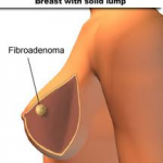
Fibroadenoma is the most common benign tumor of the breast and the most common breast tumor in women under age 30. Fibroadenomas are sometimes called breast mice or a breast mouse owing to their high mobility in the breast.
 A fibroadenoma is made up of breast gland tissue and tissue that helps support the breast gland tissue.
Black women tend to develop fibroadenomas more often and at an earlier age than white women. The cause of fibroadenomas is not known. In the male breast, fibroepithelial tumors are very rare, and are mostly phyllodes tumors. Exceptionally rare case reports exist of fibroadenomas in the male breast, however these cases may be associated with antiandrogen treatment.
Symptoms
Fibroadenomas are usually single lumps, but about 10 – 15% of women have several lumps that may affect both breasts.
Lumps may be:
- Easily moveable under the skin
- Firm
- Painless
- Rubbery
They have smooth, well-defined borders. They may grow in size, especially during pregnancy. Fibroadenomas often get smaller after menopause (if a woman is not taking hormone replacement therapy).
Exams and Tests
After a physical examination, one or both of the following tests are usually done:
- Breast ultrasound
- Mammogram
A core needle biopsy may be done to get a definite diagnosis. Women in their teens or early 20s may not need a biopsy if the lump goes away on its own or if the lump does not change over a long period of time.
Pathology
Cytology
The diagnostic findings on needle biopsy consist of abundant stromal cells, which appear as bare bipolar nuclei, throughout the aspirate; sheets of fairly uniform-size epithelial cells that are typically arranged in either an antler-like pattern or a honeycomb pattern. These epithelial sheets tend to show typical metachromatic blue staining on DiffQuick staining. Foam cells and apocrine cells may also be seen, although these are less diagnostic features.
Macroscopic
Approximately ninety percent of fibroadenomas are less than three centimetres in diameter. The vast majority of the remaining ten percent that are four centimetres or larger occur mostly in women under twenty years of age. The tumor is round or ovoid, elastic, and nodular, and has a smooth surface. The cut surface usually appears homogenous and firm, and is grey-white or tan in colour. The pericanalicular type (hard) has a whorly appearance with a complete capsule, while the intracanalicular type (soft) has an incomplete capsule.
Microscopic
Fibroadenoma of the breast is a benign tumor composed of two elements: epithelium and stroma. It is nodular and encapsulated, included in breast. The epithelial proliferation appears in a single terminal ductal unit and describes duct-like spaces surrounded by a fibroblastic stroma. Depending on the proportion and the relationship between these two components, there are two main histological features : intracanalicular and pericanalicular. Often, both types are found in the same tumor.
A) Intracanalicular fibroadenoma : stromal proliferation predominates and compresses the ducts, which are irregular, reduced to slits.
B) Pericanalicular fibroadenoma : fibrous stroma proliferates around the ductal spaces, so that they remain round or oval, on cross section. The basement membrane is intact.
Treatment
 If a biopsy shows that the lump is a fibroadenoma, the lump may be left in place or removed.
The decision to remove the lump is made by the patient and the surgeon. Reasons to have it removed include:
- Abnormal biopsy results
- Pain or other symptoms occur
- Worry or concern about cancer
They are removed with a small margin of normal breast tissue if the preoperative clinical investigations are suggestive of the diagnosis. A small amount of normal tissue must be removed in case the lesion turns out to be a phyllodes tumour on microscopic examination.
If the lump is not removed, your health care provider will watch to see if it changes or grows. This may be done using
- Mammogram
- Physical examination
- Ultrasound
A growth rate of less than sixteen percent per month in women under fifty years of age, and a growth rate of less than thirteen percent per month in women over fifty years of age have been published as safe growth rates for continued non-operative treatment and clinical observation.
Sometimes, the lump may be destroyed without removing it, using freezing. This is called cryoablation. In the procedure, ultrasound imaging is used to guide a probe into the mass of breast tissue. Extremely cold temperatures are then used to destroy the abnormal cells, and over time the cells are reabsorbed into the body. The procedure can be performed in an office setting with local anesthesia only, and leaves substantially less scarring than open surgical procedures.
The American Society of Breast Surgeons recommends the following criteria to establish a patient as a candidate for cryoablation of a fibroadenoma:
- The lesion must be sonographically visible.
- The diagnosis of fibroadenoma must be confirmed histologically.
- Lesions should be less than 4 cm in diameter.
Outlook (Prognosis)
Women with fibroadenoma have a slightly higher risk of breast cancer later in life. Lumps that are not removed should be checked regularly by physical exams and imaging tests, following the doctor’s recommendations.

